By Helen Millson (M.Phil.Sports Physio UCT)
Well-known FIFA sports physio Mario Bizzini called groin pain “The Bermuda Triangle of Sports Medicine?” (1) with good reason! There is little consensus on groin pain management. The key is the Correct Diagnosis – Easier said than done! This blog introduces key issues for more junior sports clinicians.
What is Groin Pain?
Undergraduate training often fails to emphasise the two joints in the pelvis – the hip joint and also the pubic symphysis are at the centre of many movements. (2) As a clinician, try to assess how the patient’s functional movement influences both the hip and the pubic symphysis. What causes pain and where? Try to understand the entire kinetic chain with its related function to the pelvis / groin / hip. Then perform relevant clinical tests as well as sports-specific functional tests.
The cause of groin pain is a ‘million dollar question’…….
The diagnosis is mostly by exclusion not inclusion. Osteitis Pubis (OP) diagnosis is no longer an accepted term. This may be a normal response to overload and may lead to bone stress reaction, and then possibly joint and disc degeneration. Increased signal of bone marrow oedema may be a precursor to the development of groin pain. It may or may not correlate with clinical symptoms. (3) As a clinician, consider prevention and reducing load when there are early groin pain symptoms or radiological changes (MRI) of excess load.
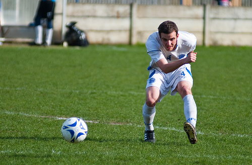
And what of the Adductor muscles? (4)
In soccer players with groin pathology, adductor dysfunction is a more frequent MRI finding than “osteitis pubis”. Both entities are mechanically related and frequently coexist.
Specific strengthening of all Adductor muscles is one of the main goals for preventing and managing groin / hip pathologies.
However, one must not be single minded and should take into account other structures including Ligamentum Teres tears (5), Ilio Psoas (6), Greater Trochanter (7), Rectus Abdominus (8), Pelvic floor muscles (9) and Gluteus muscle function (10).
Then consider Radiologists with their preferred way of imaging.
Although radiological investigations are important, most studies tend to agree that experienced clinical judgment remains a critical element in the diagnostic pathway. (11)
A few groin / hip tests have been well documented and are valuable for diagnosis. (12)
What about the treatment options?
There is consensus in the literature that non- surgical treatment should always be applied before surgery is considered. However, the time span differs in the studies.
The rehabilitation protocols show gradual progression based on objective functional and clinical markers over a reasonable time frame.
In my experience, it is of value to always have pre-season specific baseline tests (Musculo-Skeletal evaluation).
The rehabilitation can take anything from 3 – 12 weeks depending on the actual diagnosis e.g. if it is an overload problem, one would “actively rest” the athlete until they are able to fully function symptom free in their respective sport.
One should address the local strength first, followed by functional strength with the entire global perspective taken into account.
Reassessment criteria to judge progress and assess next level of activity with objective markers are essential in order to increase the rehabilitation appropriately.
Of course, at an elite sports level, one is mostly not given this time-frame to do conservative rehabilitation!
Post-operative rehabilitation programmes varies from 10 days – 12 weeks.
This depends on the type of surgery, the specific demands and………the very different requirements of the Surgeon involved!
And surgery:
• No consensus as to an ideal operating technique
• Serial patient outcome measurements are needed to base intervention success on factors other than return to sports activity.
• Operate on asymptomatic side, as it has been suggested that 40% progress to bilateral?
There is also on-going controversy regarding the prevalence of a True Hernia, with many different surgical implications. (13)
Then on to the discussion regarding hips…….
The prevalence of radiographic hip abnormalities in elite soccer players is considerable. (15)
One must identify the relationship between these radiographic abnormalities and the clinically symptomatic pathologies.
A battery of tests should be utilised to improve the accuracy of the clinical reasoning.
Hip joint restriction often precedes the development of chronic groin injury and may be a risk factor for this condition.
One must also remember that the Acetabular Labrum and Ilio-Femoral Ligament are vital for normal hip mechanics and excessive removal of either in surgery can be detrimental. (16)
As our understanding of FAI and chondral injuries and their causes grows, future efforts will focus on prevention.
Future research is required to determine the extent to which physio intervention aimed at improving hip kinematics would be effective in treating individuals with labral injuries
CONCLUSION
• The challenge lies between ascertaining the Anatomical diagnosis vs. Pathological diagnosis vs. Functional diagnosis – the interaction of the three will influence prognosis and management, whereas identification of one alone will give a bias in one direction.
SOLUTION?
• In spite of minimal EBM, it seems the most pertinent point is that many of the groin /hip pathologies can be averted by thorough and specific pre-habilitation, bearing in mind the entire kinetic chain and addressing total function around the pelvis above and below.
References
1) Bizzini M. “Warm: Up the Bermuda Triangle of Sports Medicine?” in BJSM 2011.
2) William Meyer, FA Conference London Dec 2011
3) Paajanen H. 2009. “Sports hernia” and osteitis pubis in an athlete. Duodecim. 125(3):261-6.
4) Wiktorsson-Möller, Oberg , Ekstrand , Gillquist,1983. AJSM; Lynch SA, Renström PA.1999. Sports Med; Orchard et al., 2005. Clin J Sport Med; Cunningham et al. 2007; Phillipon 2009; Lloyd 2009; Thorborg 200; Crow 2010; Gilmore 2011; Davis et al, 2011; Connell 2011; Schilders 2012 and many others.
5) Botser IB, Martin DE, Stout CE, Domb BG. 2011. Tears of the ligamentum teres: prevalence in hip arthroscopy using 2 classification systems. Am J Sports Med. Jul;39 Suppl:117S-25S.
6) Hölmich P. 2007. Long-standing groin pain in sportspeople falls into three primary patterns, a “clinical entity” approach: a prospective study of 207 patients. BJSM. Apr;41(4):247-52; discussion 252. Epub 2007 Jan 29.
7) Steinbrueck A, Hocke S, Heimkes B. 2011.Apophyseolysis of the greater trochanter through excessive sports: a case report. Am J Sports Med. Jan;39(1):195-8.
8) Connell D, Ali K, Javid M, Bell P, Batt M, Kemp S. 2006. Sonography and MRI of rectus abdominis muscle strain in elite tennis players. Roentgenol. AJR Am J. Dec;187(6):1457-61.
9) Ruth C. Lovegrove Jones, Qiyu Peng, Maria Stokes, Victor F. Humphrey, Christopher Payne, Christos E. Constantinou. Mechanisms of pelvic floor muscle function and the effect on the urethra during a cough. Eur Urol 2010;57:1101-10.
10) Graham RB, Costigan PA, Sadler EM, Stevenson JM. 2011. Local dynamic stability of the lifting kinematic chain. Gait Posture. Oct;34(4):561-3; Philippon MJ, Decker MJ, Giphart JE, et al.2011. Rehabilitation exercise progression for the gluteus medius muscle with consideration for iliopsoas tendinitis: an in vivo electromyography study. Am J Sports Med. Aug;39(8):1777-85. Epub 2011 May 12.
11) Garvey JF. 2011. Computed tomography scan diagnosis of occult groin hernia. Hernia. 2011 Dec 14.
12) Delahunt et al. Man Ther. 2011; Anthony Hogan, FA Conference London 2008; Pers Holmlich, BJSM 2004, BJSM 2007; Mallarias, Hogan et al BJSM 2009; James Moore Rehabilitation Chapter in Prof Haddad Book – “The Young Adult Hip in Sport”. To be published 2012.
13) Connell DG…. Patient care – crunch time. Br J Gen Pract. 2009
14) Gerhardt et al, AJSM 2011
15) Myers AJSM 2011