Keywords: anterior cruciate ligament reconstruction; anterior cruciate ligament healing; rehabilitation
Why is this study important?
Current treatment options for anterior cruciate ligament (ACL) rupture often result in unsatisfactory outcomes such as sport and activity limitations, instability and additional knee injuries, persistent pain, early onset of knee osteoarthritis, and reduced long-term quality of life. Treatments for ACL injury are based on the assumption that a ruptured ACL does not heal. Few studies have explored the ability for a ruptured ACL to heal following non-surgical management. It is also unclear if a healed ACL, as visualized on magnetic resonance imaging (MRI), translates into better outcomes for patients. Research investigating the healing capability of the ruptured ACL will help guide clinical practice and enable informed decision making about treatment of ACL injury.
How did the study go about this?
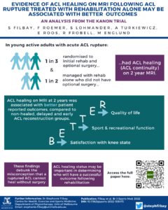
We used data from the Knee Anterior Cruciate Ligament Non Surgical versus Surgical Treatment (KANON) randomized controlled trial, the first trial to randomize young active adults with acute ACL rupture to either management with early ACL reconstruction and postoperative rehabilitation, or initial rehabilitation and a delayed ACL reconstruction if needed. The KANON trial found that clinical and structural outcomes were similar between groups at 2- and 5-year follow-up. We analyzed MRIs obtained at six time points between baseline and 5-year follow-up, to assess whether the ACL had healed after management with rehabilitation. All participants had acute ACL rupture at baseline (discontinuity of ACL fibres), and we considered a continuous ACL on follow-up MRI to represent evidence of healing (Figure 1). We also compared 2- and 5-year outcomes (including knee function, quality of life, activity level, passive knee laxity, osteoarthritis, patient acceptable symptomatic state (PASS) and treatment failure criteria) based on healing status and treatment group.
What did the study find?
Two years after injury, evidence of healing of the ruptured ACL on MRI was observed in approximately half (53%) of young adults managed with rehabilitation alone who did not cross over to delayed ACL reconstruction. One-in-three (30%) participants randomized to optional delayed ACL reconstruction had MRI evidence of ACL healing.
Participants in the healed ACL group reported better 2-year sport and recreational function and quality of life compared to the non-healed, early reconstructed and delayed reconstructed groups (between group differences were large, ranging from 11 to 28 points on the Knee Injury and Osteoarthritis Outcome Score (KOOS)). Two years after injury, a high proportion (63-94%) of participants with ACL healing on MRI were likely to feel satisfied with their level of knee pain, symptoms, function, and quality of life, compared to 29-61% in the non-healed or reconstructed groups. No one with a healed ACL reached the criteria for treatment failure, compared to 6-21% within the non-healed or reconstructed groups (Figure 2).
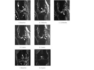
Figure 1. Evidence of ACL healing on MRI over 5 years in a KANON study participant (male, aged 31 years at the time of injury) randomised to rehabilitation and optional delayed ACLR.
- Baseline sagittal short tau inversion recovery (STIR) MRI shows complete disruption of the ACL, which is depicted as a hyperintense thickened structure (arrowhead). In addition, image depicts a characteristic traumatic bone marrow lesion (also referred to as bone contusion) in the posterior lateral tibia (arrow).
- 3-month follow-up MRI shows complete resolution of bone contusion. There is beginning scar formation with partial hypointensity in the course of the ACL. Scar is still markedly thickened.
- At 1 year, there is near-complete normalization of scar formation with a re-ligamentisation and regular course. There is some remaining intraligamentous hyperintensity (arrowhead).
- 2 years after the injury there is complete normalization of structure and signal indicating healing of the ligament.
- At 5 years persistent normalization with regular ACL course and signal intensity is depicted (arrowheads).
- Corresponding coronal STIR MRI at baseline confirms the disrupted hyperintense ACL near the proximal femoral attachment (arrowhead). There are large bone contusions at the medial and lateral tibia and lateral femur (arrowheads).
- At 5 years, normalisation with a now hypointense healed ACL, is shown also in the coronal STIR image. There is compete resolution of bone contusions.
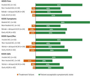
Figure 2. The percentage of participants meeting criteria for patient acceptable symptom state and treatment failure for each KOOS subscale at 2-year follow-up. Percentages do not add up to 100%, the missing percentage is explained by participants who scored above the criteria for treatment failure and below the criteria for patient acceptable symptomatic state for a given KOOS subscale.
What are the clinical implications?
This study debunks the misconception that a ruptured ACL cannot heal without surgery, while healing of ACL rupture was associated with favorable patient-reported outcomes.
Even though participants were not aware of their healing status on MRI, very few participants with ACL healing on MRI decided to undergo a delayed ACL reconstruction. This suggests the healing status of the ACL may be important in determining who will have a successful outcome following rehabilitation.
Since evidence of healing was seen on MRI as early as 3 months after ACL injury, it is possible that initial rehabilitation could reduce the number of ACL reconstructions with comparable or better outcomes for the patient. Further research is needed to determine whether the healing status of the ACL should inform treatment decisions following an initial period of rehabilitation.
When discussing ACL management options, patients should be aware of the possibility for an ACL rupture to heal after management with rehabilitation. They should also be aware that this is an emerging area of research and several questions remain unanswered, including:
- Who is most likely to experience ACL healing?
- Can specific treatments increase the likelihood of ACL healing?
- Does ACL healing (as seen on MRI) restore ACL function?
- Does ACL healing (as seen on MRI) protect against future knee injury and osteoarthritis?
Access the full paper: https://bjsm.bmj.com/content/early/2022/11/03/bjsports-2022-105473
Reference: Filbay SR, Roemer FW, Lohmander LS, et al. Evidence of ACL healing on MRI following ACL rupture treated with rehabilitation alone may be associated with better patient-reported outcomes: a secondary analysis from the KANON trial. British Journal of Sports Medicine Published Online First: 03 November 2022. doi: 10.1136/bjsports-2022-105473
Authors:
Stephanie Filbay, Frank Roemer, Stefan Lohmander, Aleksandra Turkiewicz, Ewa Roos, Richard Frobell, Martin Englund
Correspondence:
Dr Stephanie Filbay, Department of Physiotherapy, University of Melbourne, Victoria, Australia. 3010.
Email: stephanie.filbay@unimelb.edu.au
Twitter: @stephfilbay