Sport and Exercise Medicine: The UK trainee perspective (A twice-monthly series on the BJSM blog)
By Dr Thamindu Wedatilake

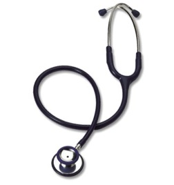
An electrician or a plumber carries a large bag, within it multiple tools aiding “diagnosis and treatment” of the household problems. Similarly doctors require tools for diagnosis and treatment of medical conditions. For a physician the tool may be a stethoscope or for a surgeon, the scalpel. A Sport and Exercise Medicine (SEM) physician primarily evaluates non-surgical musculoskeletal (MSK) problems involving muscles, tendons and ligaments. Ultrasonography (“the tool”) images these structures and their pathologies clearly.
The value of ultrasound in SEM is seen at multiple levels. Firstly ultrasound can be readily used to diagnose acute and chronic pathologies in tendons, muscles and ligaments. If it is uncertain on examination which structure is generating pain then the ultrasound probe can be placed on the area to reveal the structure that lies beneath. Ultrasound can also guide a local anaesthetic injection. Unlike a “blind” injection, US-guided injection means the clinician knows exactly which structure has been anaesthetised, and thus, helps to identify the cause of the pain. Structures can be assessed dynamically during the ultrasound scan using movement or special clinical tests; this offers important diagnostic as well as prognostic information.
Some MSK conditions require additional interventions either into a tendon, muscle, joint or ligament. The days when we blindly needled a structure and bathed the area with corticosteroid (or other substances) are slowly going. Traditionally we have referred to radiologists to perform MSK interventions. Wouldn’t it be great if you could diagnose, treat and discharge patients on the same visit!
So you have taken a history, examined and given the athlete a diagnosis and reassured them that it is safe to restart their livelihood (i.e training and competing). Is the athlete really reassured? There is vast power in visual ultrasound imges: the athlete can be reassured when shown the symptomatic structure on one side demonstrating the same pattern and findings to the corresponding structure on the contralateral side.
The act of performing an ultrasound scan is a fully interactive process that builds great rapport with the athlete. Images can be shown during the process to the athlete and also the exact area (or multiple areas) of symptoms can be fully evaluated. Furthermore it is likely that a SEM physician with ultrasound skills is more valuable to a sports team for both diagnosis and therapy using this tool.
It is important that all doctors using ultrasound work alongside (or have access to) experienced MSK radiologists so that cases can be discussed and more complex cases can be referred to radiologists for more skilled scanning and interventions.
Many clinicians working in obstetrics, emergency medicine and anaesthesia have training in ultrasonography. In the future it is likely that ultrasonography would be a compulsory part of a SEM physicians training.
So, has the time come for SEM physicians to swap their stethoscopes for an ultrasound probe?
Related BJSM Publications
AMSSM Curriculum:
- Musculoskeletal ultrasound education for sports medicine fellows: a suggested/potential curriculum by the American Medical Society for Sports by Jonathan Finnoff, Mark E Lavallee, Jay Smith
Article
Special issue:
Podcasts:
- Musculoskeletal ultrasound with Kim Harmon and Sean Martin
- What is the future in sports imaging? with Bruce Forster, David Hancock and John Orchard
Blog:
***************************************************************
Dr Thamindu Wedatilake is a Sport and Exercise Medicine registrar in the Oxford Deanery. He teaches on postgraduate musculoskeletal ultrasound courses at Bournemouth University.
Dr James Thing co-ordinates “Sport and Exercise Medicine: The UK trainee perspective” — a twice-monthly blog.