Introduction
Meniscal tears are amongst the most common injuries seen in the sports medicine and MSK clinic affecting both athletes and active patients. In the context of long-term knee health, sports participation and return to play, “not all tears are equal” with meniscal root tears often slipping under the radar of clinicians, leaving patients at risk of joint and cartilage degeneration, functional instability, and early-onset osteoarthritis (OA).
We discuss the key clinical features and the best imaging modalities to detect a meniscal root injury, as well as the evidence for surgical meniscus-sparing treatments, and knee joint and cartilage preservation techniques to ensure the best care and recovery for patients.
Getting to the root of meniscus tears
Meniscal root tears are thought to account for approximately 10–21% of all meniscal tears (1). They can be a challenge to identify in sports medicine as clinical signs can be subtle, and they may overlap in presentation with other tear types (peripheral or main body) that can be treated with natural healing or conservative management over time. If missed, the root tear can lead to prolonged rehabilitation and longer return to play for athletes.
Causes:
- Medial = Degenerative (tendency to be degenerate)
- Lateral = Traumatic (tendency to be traumatic)
However, the distinction between lateral and medial meniscus root tears is rarely so simple. In clinical practice, older patients with medial root tears may present following a relatively innocuous movement or twist that leads to a sudden, severe, sharp and disabling pain that restricts mobility. Younger patients usually present with lateral root tears following traumatic or significant pivoting episodes which are frequently associated with concurrent anterior cruciate ligament (ACL) injuries. In both cases, a meaningful clinical examination of the knee and especially the meniscus can be difficult due to pain, swelling and restricted range of motion. Treatment decisions should therefore be guided by clinical history, cause (traumatic vs. degenerative) and radiological evidence rather than strictly by laterality.
What makes “root tears” different?
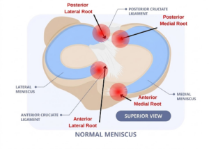
Figure 1: Superior view of the knee joint demonstrating the four primary meniscal root attachment points (highlighted in red) (2)
The roots of the meniscus have multiple roles. They help to:
- Anchor the meniscus
- Add functional stability to the meniscus with loading
- Keep the meniscus anatomically positioned
- Maintain the movement and function of the meniscus
- Maintain meniscal integrity
- Distribute load and reduce contact forces in the joint
For young athletes and active individuals, the window for intervention is narrow and delayed diagnosis or deferred treatment increases the risk of cumulative cartilage damage that can lead to early-onset OA and joint degeneration. Understanding how to detect these tears clinically and radiographically, as well as indications for orthopaedic and cartilage-sparing therapies, is key to getting your athletes back to play safely while protecting their long-term knee health.
What is a Meniscal Root Tear?
A meniscal root tear (MRT) is defined as a complete radial tear within 1 cm of the tibial meniscal attachment, or a bony avulsion at the insertion site (3).
The meniscus has four roots — anterior and posterior on both medial and lateral sides — anchoring it to the tibial plateau and maintaining hoop stress transmission (1,3).
Disrupting a root is bio-mechanically equivalent to a total meniscectomy: it abolishes hoop stresses, reduces tibio-femoral contact area, and dramatically increases compartment contact pressures (3,4).
If untreated, this leads to rapid cartilage loss and early-onset osteoarthritis (3,4).
MRTs are significantly underdiagnosed — and historically undertreated.
Who does it impact?
Two distinct populations:
Medial posterior root tears (MMPRT) — degenerative pattern
- Predominantly middle-aged women; mean age ~58 years (range 39–78) (5)
- Strong female sex predominance: up to ~93% female in some series (5)
- Accounts for 10–21% of all medial meniscal tears (4)
- Usually no history of trauma but sometimes a minor twisting event — insidious onset (6)
Lateral posterior root tears (LMPRT) — traumatic pattern
- Younger patients, typically 20–40 years (7)
- Male predominance (trauma and sport-driven) (7)
- Frequently associated with ACL tears or multi-ligament injury (8)
Presentation Pattern
- Posterior knee pain is the cardinal symptom and in an acute tear, the pain is out of proportion with typical meniscus tear patterns!
- Classic clinical clue: an audible/felt pop during low-energy activities — rising from a chair, climbing stairs, squatting
- Preceding dull popliteal or calf discomfort is common and often misdiagnosed as sciatica (10).
- No history of significant trauma in the majority of MMPRT (~70% occur in degenerative knees) (4)
- Duration from symptom onset to diagnosis: often weeks to months (6).
- On examination: posteromedial joint line tenderness; pain on forced deep flexion (7).
- Specificity of clinical examination alone is very limited — imaging with MRI is essential for diagnosis (8).
- Grading meniscal root tears is extremely valuable with the LaPrade being used to classify medial and lateral posterior root tears based on tear morphology. However, surgeons may not routinely ‘grade’ tears when considering an intervention (9)
- Acute traumatic root tears should be referred early for specialist assessment. Although earlier repair (within weeks) is certainly preferable in all patients with acute traumatic tears, successful repair can still be considered in selected chronic tears for up to 12 months post injury, depending on cartilage status, meniscal extrusion, alignment and overall joint condition.

Figure 2: Common mechanism of injuries between younger and older age groups that lead to meniscal root tears
Table 1: Risk Factors for Meniscal Root Tears
| Category | Risk Factor | Additional Information |
|---|---|---|
| Intrinsic — Non-modifiable | Older age | Mean presentation ~58 years |
| Female sex | Up to 5.9× increased risk vs male | |
| Varus mechanical alignment | 3.3× increased risk; increases medial compartment load | |
| Steep medial tibial slope | Associated with bilateral MMPRT | |
| Pre-existing OA (higher KL grade) | Often both cause and consequence | |
| Medial meniscus extrusion | Independent predictor, even after adjusting for age | |
| Ligamentous laxity | Alters load distribution | |
| Intrinsic — Modifiable | High BMI / obesity | BMI >30 confers ~4.9× increased risk |
| Sedentary lifestyle / low sports activity level | ~43% of MMPRT patients had zero recreational activity | |
| Extrinsic | ACL tear / multi-ligament injury | Primary driver for lateral root tears |
| Deep squatting / hyperflexion activities | Particularly high-load flexion >90° | |
| Iatrogenic (surgical) | Tibial tunnel reaming, tibial nailing can disrupt anterior roots | |
| High-impact sports | Pivot sports, skiing — mainly lateral root pattern |
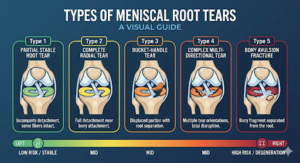
Figure 3: A visual guide of the meniscal root tears based on the LaPrade classification system (9,42)
Mechanisms, Biomechanics and Who Gets These Tears?
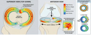
Figure 4: An infographic illustrating the biomechanical mechanism and stress distribution of transverse hoop stresses on the meniscus root within the knee joint (41).
The menisci are key to distributing axial loads through the knee via transverse hoop stresses (Figure 2) (12). If the anchoring roots are disrupted, it has a similar impact to “cutting a boat anchor”, where the supporting structures lose their ability to take on tension and distribute force on loading.
The meniscus roots are functional structures within the knee, key to effectively dispersing axial tibiofemoral loads as hoop stresses. When the roots are intact, 50–70% of the lateral and medial compartment loads are absorbed by the meniscus. When this mechanism fails, peak contact pressure can increase by 25% compared to an intact knee, equivalent to a total meniscectomy. Yang et al. (2023) demonstrated that medial meniscus posterior tears significantly increase stress across the knee joint during dynamic gait, meaning the damage is not just static but accumulates with every step and sporting movement (13).
The mechanisms of injury depend on patient profile:
Older patients
- Tend to develop chronically
- Lower energy degenerative mechanisms e.g. squatting/kneeling
- More likely to be medial meniscus
- Risk of progression to Spontaneous Insufficiency Fracture of the Knee (SIFK)
Younger patients
- Rotatory blow to the flexed knee
- Often during sport and alongside concomitant ligamentous injuries
- Present with ACL tears in up to 14–15% (10)
- More likely to be lateral meniscus
- Higher energy injuries
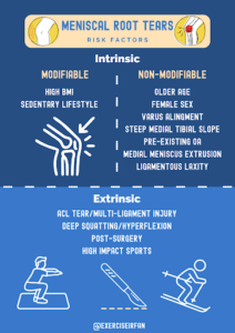
Figure 5: Modifiable and Non-Modifiable Risk Factors contributing to Meniscal Root Tears
For any sports clinician, the practical questions are always:
- What are the risks if we miss it? (8)
- How long is the athlete out for? (8)
- What is the metabolic potential for healing? (8)
- What are the sporting demands for this athlete (pivoting, cutting, player-on-player contact)? (8)
The road to recovery — what does the data in sport show?
The answer is longer than you think, and the consequences of delayed diagnosis and treatment are serious. Studies of meniscal repair in athletes show that 85% return to their previous sport at the same level, but the average time to return to sport is 6.5 months, with some elite athletes taking up to 10 months to return to their pre-operative level (8). For a root-specific repair, timelines are likely at the longer end of this range given the complexity of the fixation and the strict weight-bearing restrictions in the early post-operative phase.
It is important to emphasise that any return-to-play decision should be criterion-based rather than simply time-based, particularly where ACL reconstruction or other concomitant procedures have also been performed. This enables the patient to safely return to play and avoid aggravating other potential lingering injuries.
Sports data
A matched cohort analysis of 250 elite European soccer players across the Premier League, Bundesliga, La Liga, Ligue 1 and Serie A found a median absence of 57.5 days (equivalent to missing around 7 games) with a return to play rate of just 70%. Age over 30 was a significant negative predictor for return to play (9).
Knee injuries are relatively common in tennis, comprising around one fifth of all tennis injuries, with meniscus injuries and degenerative cartilage problems being the most prevalent in middle-aged and recreational players (11). A 10-year epidemiological study found that medial meniscus injury was specifically associated with tennis and jogging among the sports studied, reinforcing the medial compartment loading pattern seen in the sport (12). Knee injuries were among the most prevalent injury sites in both male and female professional tennis players across ATP and WTA tours between 2011 and 2016 (11). No tennis-specific meniscal root tear return-to-sport time data currently exists in the literature.
Clinical Diagnosis
Table 2: Key Clinical History Features
Domain |
Key Questions / Features |
Clinical Notes |
Mechanism of injury (8) |
Low-energy, deep flexion + axial load
e.g. squatting, rising from chair |
Degenerative MMPRT pattern |
| Twisting injury | Often with concomitant ACL rupture — LMPRT pattern | |
| “Pop” or acute onset pain | Less consistent than ligament injury | |
Symptom profile (9) |
Posterior knee pain | Particularly medial posterior joint line |
| Pain with deep flexion | Kneeling, squatting | |
| Rapid functional decline | After seemingly minor event | |
| Mechanical symptoms often absent or mild | Locking/catching less reliable than in bucket-handle tears | |
| Subtle onset | More subtle presentation than classic meniscal tears | |
Functional limitations |
Difficulty squatting / kneeling | |
| Difficulty with stair descent | ||
| Difficulty pivoting | ||
| Reduced weight-bearing tolerance | Progressive over time | |
Effusion and time course |
Subacute effusion common | |
| Acute-on-chronic or insidious worsening | Degenerative MMPRT may worsen after minor insult | |
Risk factors |
Age >40 | Mean presentation ~58 years |
| Elevated BMI | BMI >30 confers ~4.9× increased risk | |
| Varus alignment | Medial root — 3.3× increased risk | |
| Concomitant ACL injury | Lateral root — primary driver | |
| Pre-existing osteoarthritis | Often both cause and consequence | |
Red flags |
Sudden onset pain with minimal trauma | Suggests acute root avulsion |
| Posterior horn localisation | ||
| Rapid deterioration in function | ||
| Failure to improve with conservative management | Warrants urgent MRI and orthopaedic review |
Table 3: Key Clinical Tests – Sensitivity & Specificity
Note: Sensitivity and specificity data are limited for root-specific pathology. Where data are derived from general meniscal pathology cohorts, this is indicated in the Notes column.
| Clinical Test | Description | Sensitivity | Specificity | Notes |
| Painful popping event (10) | Patient-reported audible or felt pop during low-energy activity | 35% | 99.5% | High specificity, if present, strongly suggestive of root tear |
| Joint Line Tenderness (11) | Particularly posterior joint line tenderness on palpation | 44–62% | 78–90% | Moderate overall; posterior localisation increases specificity |
| McMurray’s Test (12) | Rotation with flexion/extension to provoke a click or pain at the joint line | Not established for root tears | Not established for root tears | Positive in only 57.1% of MMPRTs. Values in literature derived from general meniscal pathology, not isolated root tears |
| Akmese Sign (13) | Severe medial joint-line tenderness in near extension; minimal or no tenderness in hyperflexion with slight varus position | 86.1% | 99.1% | Best available root-specific test; high sensitivity and specificity |
| Thessaly’s Test | Weight-bearing knee rotation at 20° flexion | — | — | No root-specific data available |
| Apley’s Test | Prone compression and distraction with knee at 90° flexion | — | — | No root-specific data available |
It is important to note that although clinical tests can be effective, often the knee is simply too swollen and irritable with restricted range of motion. Therefore, provocative meniscal tests add relatively little clinical insight when considering the treatment goal and ongoing pain. This is where diagnostic imaging plays a critical role in investigating the meniscus.
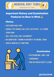
Figure 6: Summary of Patient History and Clinical Examination Findings for Meniscal Root Tears
The Role of Diagnostic Imaging
Key Imaging Findings – MRI & Ultrasound
MRI
MRI is the gold standard imaging modality for the assessment of meniscal tears. Proton density (PD) and proton density fat suppressed (PDFS) sequences are primarily used for identifying meniscal tears on MRI. Medial posterior root tears tend to be more straightforward to identify than lateral counterparts, which can mimic the appearance of standard radial tears (14).
The menisci can be assessed all three standard plains (axial, coronal and sagittal) to identify direct and indirect signs of meniscus root tears:
- Truncation / Cleft sign (coronal) — a vertical fluid signal on coronal sequences at the root attachment (15)
- Radial tear sign (axial) — axial views for tears are underused in practice but are powerful; particularly helpful when coronal signs are subtle (15)
- Ghost sign (sagittal) — high or intermediate meniscal signal due to a meniscus tear extending through the whole meniscus
- Meniscal extrusion (coronal) — indirect sign; meniscus displaced beyond the tibial margin (16)
Point of care Ultrasound – a quick tool to help you evaluate the meniscus in clinic
Point of care ultrasound – can help to identify key features of a “non-functioning meniscus”. The most obvious is meniscus extrusion at the joint line alongside secondary features.
- Joint effusion,
- increased medial meniscal extrusion – (standing, 0 degrees and 90 degrees flexion)
- Synovial hypertrophy
- Doppler signal at tib / fib (17)
If the meniscus is no longer functioning, meniscal extrusion does not decrease between extension and knee flexion positions and allows for dynamic assessment in clinic (17).
In the hands of an experienced operator ultrasound may help to provide clinicians with a higher pre test probability (good sensitivity and specificity) – and be used to screen patients who may need further MRI evaluation (18).
Table 4: A Systematic Review of MRI Signs in Meniscal Root Tears and their Diagnostic Accuracy
| Characteristic MRI Finding | Sensitivity (%) | Specificity (%) |
|---|---|---|
| Radial tear 19-22 | 67.6–94 | 94–100 |
| Truncation sign 19 | 90.0 | 100 |
| Cleft/truncation sign 23 | 56.3 | — |
| Cleft sign 22,24 | 65.6 | 95.5 |
| Truncated triangle sign 24 | 59.4 | 94.0 |
| Ghost sign19,20,22-24 | 34.4–100 | 96.7–97 |
| >3mm medial meniscus extrusion19,23,25 | 54–74.5 | 90.0–98.0 |
| Giraffe neck sign 23,26 | 81.7–87.3 | — |
| Shiny corner lesion 22,23,27 | 61.8–62.0 | 97.0 |
| Cleft + ghost 24 | 71.9 | 93.5 |
| Ghost + truncated triangle 24 | 81.3 | 91.5 |
| Cleft + truncated triangle 24 | 78.1 | 92.4 |
There are numerous MRI findings indicative of meniscal root tears which demonstrate variable levels of diagnostic accuracy. Signs such as radial tears and truncation signs exhibit high sensitivity and specificity, suggesting they are reliable indicators of meniscal root disruption. Similarly, the ghost sign and medial meniscus extrusion (>3 mm) show high specificity, but a wide range of sensitivities and their presence can help confirm a diagnosis, but absence should not be used to rule out disease.
Emerging signs such as the giraffe neck sign and the shiny corner lesion demonstrate reasonable sensitivity and high specificity, supporting diagnosis. A key consideration is that utilising these MRI findings in combination leads to greater diagnostic accuracy compared to individual findings. Combinations consistently demonstrate higher sensitivity and specificity together.
In clinical practice, the MRI helps to confirm the diagnosis when there is a strong clinical suspicion of a MRT.
MRI
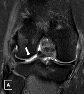
Figure A: Cleft sign in coronal PDFS (proton density fat saturated sequence). A large fluid filled cleft within the posterior root of the lateral meniscus is seen. It indicates a detachment at the root and loss of the normal shock absorbing function of the meniscus.
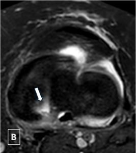
Figure B: Radial tear sign in axial PDFS. The image demonstrates an absent meniscus due to a tear at the posterior root of the medial meniscus.
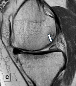
Figure C: Ghost sign in sagittal PD. There is an abnormal high/intermediate signal of the posterior horn of the medial meniscus due to partial volume artefact. This is due to a radial tear extending the full width of the meniscus parallel to the axis of the MRI slice (28).
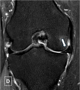
Figure D: Meniscus extrusion sign in coronal PDFS. A meniscal extrusion (>3mm) beyond the tibial articular surface, an indirect sign of a meniscal tear, here it is causing buckling of the medial collateral ligament.
Surgical Treatment Options
- Treatment decisions for meniscal root tears should be guided by the aetiology of injury and patient characteristics
- Treatment options included non-surgical management, partial meniscectomy and meniscal root repair (29)
- It is recommended that all acute root tears initially receive surgical consultation
- Meniscal root repair is the preferred treatment option as it can lead to improved clinical outcomes and reduce the risk of future knee replacement (30)
- Following surgery, rehabilitation should include restricting weight bearing, oedema control and isometric quadriceps exercises with a gradual return to impact and sports taking a minimum of 6 months (29)
A surgical consultation may not simply rely on radiological grading to determine treatment planning. The most important questions to ask are:
- Is this a complete or functionally significant root tear?
- Is it acute or chronic?
- Is there meniscal extrusion, and how much?
- What is the condition of the cartilage?
- What is the mechanical alignment of the knee?
- Is there an associated ligament injury?
Partial tears with preserved functional root attachment are clearly a different entity and the vast majority do not require surgical intervention.
| Procedure | Description | Indication |
| Transtibial pullout repair | Involves passing sutures through tunnels in the meniscus and securing them with a button or anchor [31] | Most common repair method [30] |
| Suture anchor repair | An anchor is inserted into the tibia which acts to secure sutures that pass through the meniscus [31] | Alternative repair technique that avoids tibial bone tunnels and the need for distal fixation [29] |
| Partial meniscectomy | Partial removal of the meniscus | Used in patients with both arthritis and chronic root tears or those with partial tears where the majority of the root attachment remains intact [31] |
| Meniscus Centralisation | A centralisation suture is attached to a knotted or knotless anchor [31] | Performed in addition to root repair and can be particularly beneficial when there is substantial meniscal extrusion [30] |
| High tibial osteotomy | Involves cutting into the tibia and creating a gap to realign the lower leg [32] | Can be considered for chronic medial root tears with varus malalignment [33] |
| Non-operative management | Analgesia, anti-inflammatory medication, physiotherapy and modification of weight-bearing activity. [29,30] | Patients with advanced osteoarthritis, severe cartilage damage or significant medical comorbidities [29,30] |
Table 5: What do you do when you see a meniscal root tear and underlying osteochondral defect?
| Size | Surgical Intervention | |
|---|---|---|
| Pure Cartilage | ||
| 1cm | Minced Cartilage Implantation | |
| 1-2cms | Minced Cartilage Implantation or Autologous matrix-induced chondrogenesis (AMIC) | |
| >2 cm² | Autologous Chondrocyte Implantation (ACI) | |
| Cartilage and Bone | ||
| 1cm | Osteochondral Autologous Transfer Surgery (OATS) | |
| 1-2cms | Mosaicplasty | |
| >2 cm² | Osteochondral allograft | |
Physiotherapy Rehabilitation Post Meniscus Root Repair
Rehabilitation following meniscus root repair is fundamentally different to the standard meniscus repair and must account for the biomechanical considerations unique to root tears (34). Post-operative physiotherapy commences immediately after surgery and progresses through four distinct phases (34,35).
- Phase One – Recovery
- Phase Two – Transition (to weight bearing)
- Phase Three – Rebuild
- Phase Four – Restore
Post-operative precautions aim to minimise joint loading or movements that contribute to excessive displacement or distraction of the repair. Generally patients follow a longer, and more cautious return to loading that with traditional tears due to the loss of the hoop stress mechanism (34). Rehab therefore must be personalised.
Early Recovery Phase (0-6 Weeks)
- NWB with knee braced in extension for ambulation — protects repair and minimises hoop stress on the root (34,36).
- Flexion restricted to 90° for the first 2 weeks, gradually progressing; full extension achieved and maintained from day one (32,33).
- Early quadriceps activation priority — SLR, isometric contractions, NMES, BFR to limit muscle inhibition and atrophy (34,35).
- Resisted hamstring exercises avoided — semimembranosus attachment risks traction on repair site (34,35,36).
Transition Phase (Weeks 6-12)
- Gradual weight bearing progression over 2–4 weeks; step count monitored via wearable tech to avoid load spikes (34,35,36).
- Shallow squatting (≤70°) from weeks 9–10; resisted hamstring curling restricted until week 16 (34,35).
- Varus alignment patients require medial unloader brace up to 16 weeks — varus is a strong predictor of long-term repair failure (34,36).
Rebuild and Restore Phase (3-12 Months)
- Progressive strengthening towards quadriceps symmetry is the priority — increased quadriceps strength following root repair is directly associated with reduced meniscal extrusion progression (34).
- Quadriceps LSI of at least 70% required before return to running, progressing to 90% before return to sport (34,35,36).
- Hop testing battery used to assess functional symmetry prior to return to impact/sport (34).
- Return to running not considered before 6 months post-op; return to sport guided by strength symmetry and hop testing criteria (34).
Return to Run/Sport
The evidence base for return to running and sport for patients with Medial posterior meniscus root tear remains limited because the typical patient profile is older with degenerative joint changes, where returning to running or sport is not always the primary goal (34,35). However, for patients with a posterior lateral root repair, which tend to occur in active younger patients, often in the context of an ACL injury, the picture is different where return to sport is expected and targeted (34,35,38).
Objective criteria must be met before return to running including, full pain free ROM, absence of joint effusion and quadriceps strength symmetry. The criteria is largely drawn from the ACL reconstruction literature given the lack of meniscus root specific RTS data (34,35,36). Given the limited evidence, return to running and sport decisions should never be made by time alone. A shared decision-making approach between the patient, physiotherapist and surgeon remains the gold standard (34,35,38).
Outcomes
The evidence for repair over non-operative management or meniscectomy is compelling. Post-op patient reported outcomes improve significantly from baseline following repair are superior to those undergoing meniscectomy (36,37). Conversion to a Total Knee Replacement is substantially lower following repair, 9.8% compared to 36% following meniscectomy at a mid and long term follow up. Patients also have a reduction of 83% reduction in the likelihood of OA progression at a minimum four year follow up (36,37).
Table 6: Post-operative Rehabilitation Protocol from Injury to Return to Play (RTP)
| Phase | Timeframe | Weight Bearing | Knee Flexion ROM | Key exercises | Precautions |
| Phase 1: Recovery | Weeks 0-6 | NWB: Knee braced in extension for ambulation | Limited to 90 degrees for 2 weeks, gradual progression thereafter | Static quad, SLR, isometric contractions, NMES, BFR, passive ROM. | No resisted hamstrings, no squatting, full extension from day one. |
| Phase 2: Transition | Weeks 6-12 | Gradual WB progression to FWB | Progress forwards fill symmetrical ROM | Hip hinging, OKC quad strengthening, shallow squats < 70 degrees from 9-10 weeks | No hamstring curling until week 16. No rotation or cross- legged sitting |
| Phase 3: Rebuild | 3-6 Months | Full WB | Full ROM | Progressive quad and lower limb strengthening, squat depth progression, functional loading | Quad LSI >70% required before returning to running. |
| Phase 4: Restore | 6-12 Months | Full WB | Full ROM | Sport specific loading, return to running, hop testing | RTS criteria: Quad LSI > 90%, hop testing, minimal joint irritation and no return to running before 6 months post op. |
Metabolic Challenges with Meniscal Injuries
Subchondral insufficiency fractures of the knee (SIFK) are a challenging injury to deal with, as they often co present with meniscal pathology of the knee. They are thought to be due to a combination of:
- Mechanical insult due to a de-functioning meniscus tear
- Metabolic predisposition (Menopause status)
- Bone marrow oedema syndrome
- Excess bodyweight (BMI>30)
- Low bone mineral density (osteopenia) (39)
SIFK acts like a metabolic chameleon hiding alongside meniscal injuries, and should be considered as a part of the work up for meniscal root tears. In one study, SIFK was reported to be present in 3% of Middle Ages patients with symptomatic (knee pain) and suspected meniscal tears (40). If undetected, SIFK can lead to rapid joint degeneration and conversion to surgery/joint replacement in the medium term.
Early identification of this injury can help to minimise loading of the symptomatic bone and aim to preserve joint function by a period of protected weight bearing and bone health optimisation. In practical terms many of these patients will be under shared care with Orthopaedic Surgeons and Sports Physician/Rheumatologists for metabolic health screening and optimisation.
Many surgical interventions are reserved for when the acute osseous process in SIFK settles rather than immediately operating on a very irritable knee. This period is crucial to allow for protected loading, reassessment of symptoms and appropriate investigation/optimisation of bone health where indicated. This is particularly important in the older female patient population. Only when this acute process settles can there be a discussion of undertaking definitive root surgery.
Table 7: Diagnostic Tips for SIFK in Meniscal Root Tears
| Key Steps | Investigation | How will it help |
| Detect the injury early | MRI scan of the knee | Identify bone marrow oedema early, and the site :
(Tibial v Femoral) (Medial v Lateral) This will help to identify patients who need to be offloaded for 6-8 weeks acutely. |
| Screen for metabolic health | • Urea and Electrolytes (U&Es)
• Liver Function Tests (LFTs) • Calcium / corrected calcium • Phosphate • Vitamin D (25-OH) • Full blood count • Thyroid function test • HBA1c |
Identify treatable and correctable deficiencies that can impact bone health. |
| Bone density | DEXA Scan | Identify patients at risk of osteoporosis |
Conclusion
In conclusion, there are four key take home messages when it comes to Meniscal Root Tears:
- Meniscus root tears are severe, requiring early and urgent diagnosis to preserve knee function.
- In patients where there is a strong clinical suspicion of a meniscal root tear, MRI imaging should be arranged as early as possible.
- Complete meniscal root tears, increase the risk of OA, SIFK and need for surgical intervention.
- Patients diagnosed with SIFK after a root tears should undertake a metabolic bone health assessment
Authors and Affliations
- Dr Muhammad Umer , FY2 Doctor , Imperial College Healthcare NHS Trust, umer_afreen@hotmail.co.uk / Muhammad.umer7@nhs.net
- Alifiyah, 3rd Year Medical Student, King’s College London
- Aadil Master, Specialist MSK Physiotherapist, Wrightington, Wigan and Leigh NHS Foundation Trust
- Dr Tamer Ahamed, GPST1, Barking, Havering & Redbridge University Hospitals NHS Trust
- Dr Salmaan Ahmed, FY2 Doctor, Barking, Havering & Redbridge University Hospitals NHS Trust
- Dr Rifat Hassan, ST2 Radiology Registrar, Royal Free London NHS Foundation Trust
- Dr Kardo Ala-Aldeen, ST4 Radiology registrar, Luton and Dunstable Hospital
- Dr Preena Patel , Radiology Consultant, Luton and Dunstable Hospital
- Mr Kash Akhtar, Consultant Orthopaedic surgeon, Cleveland Clinic
- Dr Irfan Ahmed, Consultant in Musculoskeletal, Sport & Exercise Medicine, ‘The MSK Playbook’
References
- Lassioued, O., Amzallag, J., Abbassi, H., Fariborz, H., Naanaa, T., LASSIOUED, O., AMZALLAG, J., ABBASSI, H., FARIBORZ, H. and NAANAA, T., 2026. Meniscal Repair in Athletes: Functional Outcomes and Return to Sport. Cureus, 18(1)
- Ramkumar PN. Meniscus Root Tear [Internet]. Long Beach (CA): Prem N. Ramkumar; 2026, cited 2026 May 20]. Available from: https://ochipandknee.com/meniscus-root-tear-knee-specialist-los-angeles-orange-county-ca/
- Lavoie-Gagne, O.Z., Korrapati, A., Retzky, J., Bernstein, D.N., Diaz, C.C., Berlinberg, E.J., Forlenza, E.M., Fury, M.S., Mehta, N., O’Donnell, E.A. and Forsythe, B., 2022. Return to play and player performance after meniscal tear among elite-level European soccer players: a matched cohort analysis of injuries from 2006 to 2016. Orthopaedic Journal of Sports Medicine, 10(1), p.23259671211059541.
- Kushwaha, S., Khan, F.A., Kumar, P., Singh, S. and KUSHWAHA, S., 2023. Meniscal and chondral injury patterns in athletes with anterior cruciate ligament tear. Cureus, 15(11).
- Renström, P.A., 1995. Knee pain in tennis players. Clinics in sports medicine, 14(1), pp.163-175.
- Majewski, M., Susanne, H. and Klaus, S., 2006. Epidemiology of athletic knee injuries: a 10-year study. The knee, 13(3), pp.184-188.
- Yang, Q., Zhu, X.Y., Bao, J.Y., Zhang, J., Xue, A.Q., Wang, D.Y., Mao, Z.M., Tang, J.W., Jiang, D., Fan, Y. and Zhao, F., 2023. Medial meniscus posterior root tears and partial meniscectomy significantly increase stress in the knee joint during dynamic gait. Knee Surgery, Sports Traumatology, Arthroscopy, 31(6), pp.2289-2298.
- Pache S, Aman ZS, Kennedy M, Nakama GY, Moatshe G, Ziegler C, LaPrade RF. Meniscal Root Tears: Current Concepts Review. Arch Bone Jt Surg. 2018 Jul;6(4):250-259.
- Moatshe G, Chahla J, Slette E, Engebretsen L, Laprade RF. Posterior meniscal root injuries. Acta Orthop. 2016 Oct;87(5):452-8.
- Bae JH, Paik NH, Park GW, Yoon JR, Chae DJ, Kwon JH, Kim JI, Nha KW. Predictive value of painful popping for a posterior root tear of the medial meniscus in middle-aged to older Asian patients. Arthroscopy. 2013 Mar;29(3):545-9. doi: 10.1016/j.arthro.2012.10.026. Epub 2013 Jan 29. PMID: 23375180.
- Shekarchi B, Panahi A, Raeissadat SA, Maleki N, Nayebabbas S, Farhadi P. Comparison of Thessaly Test with Joint Line Tenderness and McMurray Test in the Diagnosis of Meniscal Tears. Malays Orthop J. 2020 Jul;14(2):94-100. doi: 10.5704/MOJ.2007.018. PMID: 32983383; PMCID: PMC7513660.
- Lee JH, Lim YJ, Kim KB, Kim KH, Song JH. Arthroscopic pullout suture repair of posterior root tear of the medial meniscus: radiographic and clinical results with a 2-year follow-up. Arthroscopy. 2009 Sep;25(9):951-8. doi: 10.1016/j.arthro.2009.03.018. PMID: 19732632.
- Garcia JR, Ayala SG, Allende F, Mameri E, Haynes M, Familiari F, Geeslin AG, Murray I, Moatshe G, Verma NN, LaPrade RF, Chahla J. Diagnosis and Treatment Strategies of Meniscus Root Tears: A Scoping Review. Orthop J Sports Med. 2024 Nov 1;12(11):23259671241283962. doi: 10.1177/23259671241283962. PMID: 39493310; PMCID: PMC11531027.
- Weerakkody Y, Al Kabbani A, Hacking C, et al. Meniscal root tear. Reference article, Radiopaedia.org https://doi.org/10.53347/rID-40036
- Choi SH, Bae S, Ji SK, Chang MJ. The MRI findings of meniscal root tear of the medial meniscus: emphasis on coronal, sagittal and axial images. Knee Surg Sports Traumatol Arthrosc. 2012 Oct;20(10):2098-2103. doi: 10.1007/s00167-011-1794-4. Epub 2011 Nov 24. PMID: 22113225.
- Hantouly, A.T., Aminake, G., Khan, A.S. et al. Meniscus root tears: state of the art. International Orthopaedics (SICOT) 48, 955–964 (2024). https://doi.org/10.1007/s00264-024-06092-w
- Nishimura M, Takata Y, Shima Y (2025). Five key ultrasound findings associated with medial meniscus posterior root tear. The Knee. 55, 186-192
- Hassan, R., Poku, D., Chan, O. et al. Diagnostic accuracy of ultrasonography for meniscal tears: a systematic review and meta-analysis. Skeletal Radiol (2025). https://doi.org/10.1007/s00256-025-05076-0
- Choi SH, Bae S, Ji SK, Chang MJ. The MRI findings of meniscal root tear of the medial meniscus: emphasis on coronal, sagittal and axial images. Knee Surgery, Sports Traumatology, Arthroscopy. 2011 Nov 24;20(10):2098–103.
- Lee YG, Shim JC, Choi YS, Kim JG, Lee GJ, Kim HK. Magnetic Resonance Imaging Findings of Surgically Proven Medial Meniscus Root Tear. Journal of Computer Assisted Tomography. 2008 May;32(3):452–7.
- Lee SY, Jee WH, Kim JM. Radial Tear of the Medial Meniscal Root: Reliability and Accuracy of MRI for Diagnosis. American Journal of Roentgenology. 2008 Jul;191(1):81–5.
- Kaneko S, Hashimoto S, Honda A, Ohsawa T, Takase R, Shimada T, et al. The Usefulness of Posterior Shiny Corner Lesions in the Early Diagnosis of Medial Meniscus Posterior Root Tears. Cureus. 2024 May 19.
- Okazaki Y, Furumatsu T, Kajiki Y, Hiranaka T, Kintaka K, Kodama Y, et al. A posterior shinycorner lesion of the tibia is observed in the early phase after medial meniscus posterior root tear. European journal of orthopaedic surgery & traumatology : orthopedie traumatologie [Internet]. 2022 Feb;32(2):301–6.
- Asai K, Nakase J, Oshima T, Shimozaki K, Toyooka K, Tsuchiya H. Lateral meniscus posterior root tear in anterior cruciate ligament injury can be detected using MRI-specific signs in combination but not individually. Knee Surgery, Sports Traumatology, Arthroscopy. 2019 Jun 28;28(10):3094–100.
- Park H-J, Kim SS, Lee S-Y, Choi Y-J, Chung E-C, Rho M-H, et al. Medial meniscal root tears and meniscal extrusion transverse length ratios on MRI. The British Journal of Radiology. 2012 Nov;85(1019):e1032–7.
- Furumatsu T, Fujii M, Kodama Y, Ozaki T. A giraffe neck sign of the medial meniscus: A characteristic finding of the medial meniscus posterior root tear on magnetic resonance imaging. Journal of Orthopaedic Science. 2017 Jul;22(4):731–6.
- Chang EY, Chen KC, Chung CB. The shiny corner of the knee: a sign of meniscal osteochondral unit dysfunction. Skeletal Radiology. 2014 Jul 23;43(10):1403–9.
- MSK MRI Helms third edition- https://doi.org/10.1016/C2013-0-18906-4
- Bhatia S, LaPrade CM, Ellman MB, LaPrade RF. Meniscal Root Tears. The American Journal of Sports Medicine. 2014 Mar 12;42(12):3016–30.
- Chahla J,et al. International Delphi Consensus on Medial Meniscal Root Tears Shows High Agreement on Diagnosis, Treatment, and Rehabilitation but Lack of Agreement on Treatment of Asymptomatic Tears. Arthroscopy: The Journal of Arthroscopic & Related Surgery. 2025 Nov;41(11):4683–98.
- Gerhold C, Dave U, Bi AS, Chahla J. Medial Meniscus Root Tears: Anatomy, Repair Options, and Outcomes. Arthroscopy : the journal of arthroscopic & related surgery : official publication of the Arthroscopy Association of North America and the International Arthroscopy Association [Internet]. 2025 Apr;41(4):871–3. Available from: https://pubmed.ncbi.nlm.nih.gov/40107834/
- Royal Orthopaedic Hospital . Royal Orthopaedic Hospital – High Tibial Osteotomy and Distal Femoral Osteotomy [Internet]. roh.nhs.uk. Available from: https://roh.nhs.uk/services-information/knees/hto-and-dfo
- Junya Itou, Umito Kuwashima, Itoh M. High tibial osteotomy for medial meniscus posterior root tears in knees with moderate varus alignment can achieve favorable clinical outcomes. Journal of Experimental Orthopaedics. 2022 Jul 7;9(1).
- Monson, J.K. and LaPrade, R.F., 2025. Posterior medial meniscus root tears: Clinical implications, surgical management, and post-operative rehabilitation considerations. International Journal of Sports Physical Therapy, 20(1), p.127.
- Kawada, K., Furumatsu, T., Fukuba, M., Tamura, M., Higashihara, N., Okazaki, Y., Yokoyama, Y., Katayama, Y., Hamada, M. and Ozaki, T., 2023. Increased quadriceps muscle strength after medial meniscus posterior root repair is associated with decreased medial meniscus extrusion progression. BMC Musculoskeletal Disorders, 24(1), p.727.
- Eseonu, K.C., Neale, J., Lyons, A. and Kluzek, S., 2022. Are outcomes of acute meniscus root tear repair better than debridement or nonoperative management? A systematic review. The American Journal of Sports Medicine, 50(11), pp.3130-3139.
- Krivicich, L.M., Kunze, K.N., Parvaresh, K.C., Jan, K., DeVinney, A., Vadhera, A., LaPrade, R.F. and Chahla, J., 2022. Comparison of long-term radiographic outcomes and rate and time for conversion to total knee arthroplasty between repair and meniscectomy for medial meniscus posterior root tears: a systematic review and meta-analysis. The American Journal of Sports Medicine, 50(7), pp.2023-2031.
- Mueller, B.T., Moulton, S.G., O’Brien, L. and LaPrade, R.F., 2016. Rehabilitation following meniscal root repair: a clinical commentary. journal of orthopaedic & sports physical therapy, 46(2), pp.104-113.
- Zoe E. Zimmerman, Leslie J. Bisson, Jeffrey N. Katz (2021). Perspective on subchondral insufficiency fracture of the knee,Osteoarthritis and Cartilage Open. 3(3), 100183.
- Jamie L. Huizinga, et al (2020). Prevalence of undiagnosed subchondral insufficiency fractures of the knee in middle age adults with knee pain and suspected meniscal tear, Osteoarthritis and Cartilage Open. 2(4), 10089
- Image generated using Adobe Firefly from the prompt ‘meniscus and knee structures showing the stress distribution of transverse hoop stresses on the meniscus root in colour’
- Image generated using Gemini from the prompt ‘grades 1-5 of meniscal root tears in the knee using colourful graphics’