Authors: Daniel M Cushman, Derek Stokes, Leyen Vu, Blake Corcoran, Michael Fredericson, Sarah F Eby, Masaru Teramoto
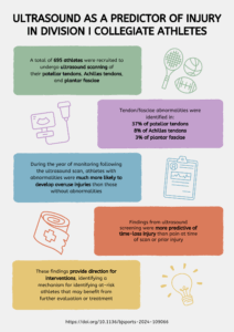
In this blog we will explain how standard B-mode ultrasound was used to identify precursors to injury in the patellar tendon, Achilles tendon, and plantar fascia in National Collegiate Athletics Association (NCAA) Division I (DI) collegiate athletes. Our study recently published in BJSM looked at 695 student-athletes at the University of Utah, Washington State University, and Stanford University.
Why is this study important?
Collegiate student-athletes are at risk of developing symptomatic patellar tendinopathy, Achilles tendinopathy, and plantar fasciopathy, which can cause time away from sport and even medical retirement. Prevention of these injuries has been challenging. While a prior history of injury and the presence of mild symptoms may help identify those at risk for time-loss injury, the association is not perfect. Ultrasound is a relatively inexpensive tool that can be used to assess tendon appearance; it has been shown in other studies that abnormal ultrasound appearance is associated with future symptom development. However, this has not been demonstrated in the collegiate population, nor has it been demonstrated to be superior to simple patient history.
How did the study go about this?
This study included 695 athletes from three institutions spanning 18 different sports. All participants completed a questionnaire asking about current and prior symptoms, as well as demographic information. Each subject had their patellar tendons, Achilles tendons, and plantar fasciae scanned in a standardized fashion, recorded as blinded videos. Later, a blinded reviewer looked at each video to identify the presence of abnormalities in these structures. The participants were then monitored for a year to see if they developed an overuse injury to any of the three structures.
What did the study find?
Abnormalities were seen in many structures (37% of patellar tendons, 8% of Achilles tendons, and 3% of plantar fasciae). Those with abnormalities were much more likely to develop injuries compared to those with abnormalities for all three structures. Furthermore, these abnormalities were more predictive of an impending time-loss injury than either a history of prior injury to the area, or pain in the area at the time of scanning. This method was not an effective screening test, however, as most subjects with abnormalities remained injury-free; only 1-in-5 to 1-in-10 of athletes with an abnormality become injured. Importantly, though, those who had normal pre-season ultrasound scans were almost completely uninjured at a year.
What are the key take-home points?
We found three main points from this study for the patellar tendons, Achilles tendons, and plantar fasciae in DI collegiate athletes:
- A simple ultrasound screening test was more predictive of time-loss injury over the upcoming year than pain at the time of scanning or prior injury.
- Those with normal tendons/fasciae were very unlikely to be injured over a year.
- Those with abnormalities were still relatively unlikely to be injured but more at-risk than those with normal tendons/fasciae.
The findings from this study will hopefully allow for interventions for at-risk athletes, likely with a thorough functional evaluation of the athlete, identifying the cause of these structural changes.