 The smell of formaldehyde will never leave me. On my first day as a medical student, in anatomy class, six of us crowded around a dead body, scalpels in hands, waiting to make the first cut. On my university entry form, like everyone else I had proudly stated that I wanted to “help people.” However idealistic my goals, I had assumed that these “people” would at least give me a sporting chance to help by being alive. On that day, it was not to be, and for three more years I consigned myself to seeing bodies in parts.
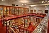 Had I been a medical student in 19th century America, however, the study of cadavers would not have been on my curriculum. Literally and figuratively, dissection was not on the table. A certain Dr Thomas Dent Mütter (1811-1859), former student at the University of Pennsylvania medical school, was to set in motion a different trend. After leaving America to train in Paris, where dead bodies were more readily available for science (and a dime a dozen), he returned to the US to become professor of what would today be known as plastic and reconstructive surgery. During his life, Mütter amassed a collection of “anatomical specimens, models, and medical instruments,” which, on his death, were bequeathed to the College of Physicians of Philadelphia, the oldest medical society in the US, which has added to and maintained the collection ever since. I visited the Mütter museum a couple of weeks ago.
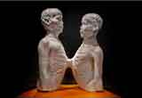 Parts again, fragmented human beings. These were my first, and not at all unpleasant, thoughts as I saw: a plaster cast of the world’s first “Siamese” (conjoined) twins, Chang and Eng Bunker, the tallest (acromegalic) skeleton in North America; a piece of the thorax of John Wilkes Booth, who assassinated President Lincoln, and a human-leather-bound book, “tanned from the skin of the thighe” of a woman called Mary.
There is something reassuring about deconstructing complex matter. Maybe we should call it “specimenology.” Maybe someone already has. The Mütter is highly visited on account of one very famous specimen, Einstein’s brain. Acquired under slightly dubious circumstances by the pathologist Thomas Harvey, who carried out the post-mortem, it was kept “sometimes in a cider box under a beer cooler.” Sure, why not. The cross-sections show a “large parietal lobe” and “no sylvan fissure.” Unsurprisingly, from these anatomical idiosyncrasies have emerged all sorts of public inferences about Einstein’s intellectual capabilities. The idea of “brain mapping” in this way reminds me of the “homunculus,” that caricature human whose distorted organs appear splayed across the brain’s surface, visually demonstrating the proportions of sensorimotor “brain space” presumed to be allocated to different body parts.
Another fragmented approach to medicine, the disease-specific one, can lead to significant achievements, particularly when certain diseases, or the devastating complications of late presentations, become consigned forever to glass cases. In one such cabinet in the Mütter resides a very large, faecally loaded colon of a man who, in the 1890s, went to his surgeon and was discharged with presumed constipation. However, the constipation was the result and not the cause of his colonic enlargement, and he died shortly thereafter, with what would now be diagnosed at Hirschsprung’s. Other afflictions remain stubbornly unresolved. For example, it is a truth universally acknowledged that a child can swallow anything under the sun. Or so would have thought ENT surgeon Chevalier Jackson if he ever browsed his collection of swallowed and rescued foreign bodies, including bolts, nails and a “perfect attendance” pin, all carefully labelled and organised in a set of drawers.
A more holistic view of illness is presented in a lovely little exhibit called “medical motifs in fairy tales,” which alludes to the art of diagnosis-by-inspection. For example, in “The Tale of the Three Spinners” the Brothers Grimm describe deformities acquired from years of sitting at a spinning wheel: a flat foot (from peddling), a pendulous lower lip (from licking), and an oversized thumb (from twisting thread).
Body parts finally unite in the “Soap Lady,” a 19th century woman whose death is shrouded in mystery and whose corpse, under certain environmental conditions, literally turned to soap, forming an “adipocere.” The soap cast lies in a glass coffin. Nobody knows her name.
This delightful museum presents the scientific thinking of the time, which can now be challenged. Additional vignettes reveal a historical context of slavery, colonialism, Orientalism and voyeuristic displays of the marginalised. To me, this all symbolises centuries of Western scientific endeavour, with its emphasis on mind-body duality, abstraction, decontextualisation, and formalisation of knowledge. The medical and public health benefits have been substantial. But what about the sacrifices? Can a viewer ever reach an understanding of these delicately excised, exposed exhibits as human beings?
The moniker “Soap Lady” reminds me of a time when I was a senior house officer during a busy hospital take. A medical student had come to join us, keen to absorb whatever teaching I could offer. During a rare lull, she turned to me and asked, “Why do we say ‘there is an MI in resus,’ or ‘an LVF in bed 4?’ ‘Why do we call patients by their diseases?'” The extraordinary simplicity of this question, the guilelessness behind it, unsettled me that day and has left me thinking ever since. Because, although it’s mostly a matter of efficiency, will this language that our profession takes for granted have unintended consequences for how future doctors regard their patients?
I absolutely do not regret my thorough grounding in basic science. Sorting, cleaving, occasionally butchering; peeling back layer after layer, to reveal a scientific “truth.” Of all the topics most clinically relevant to my practice today, anatomy would be up near the top. People like Mütter, and others around the world, helped to advance this educational agenda. However, right from that first day in the dissecting room, where altruistic dreams meet the smell of death, to now, where harangued staff administer to “the LVF in bed 4,” a power seems to exist that strives to inculcate the dehumanisation of patients into generations of medical trainees. Maybe we should take a moment now and again to remember the people behind the parts.
Photo 1: The Main Gallery of Philadelphia’s Mütter Museum George Widman, 2009, for the Mütter Museum of The College of Physicians of Philadelphia
Photo 2: Death Cast of “Siamese Twins” Chang and Eng Bunker George Widman, 2009, for the Mütter Museum of The College of Physicians of Philadelphia
Photographs reproduced with the kind permission of the College of Physicians of Philadelphia. Many thanks to Jenn Sem for introducing me to the Mutter.
Suchita Shah is a general practitioner in Oxford, UK. She is currently living in Boston and is a student at the Harvard School of Public Health, Boston.