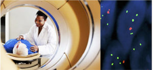
In the current issue of JNNP, Iwadate and colleagues investigated the correlation between the changes in 11C-methionine positron emission tomography (PET) and the 1p/19q status in oligodendroglial tumours.
Gliomas are the most common primary brain tumour in adults. Typically, based in the histological classification, gliomas have been classified into two major subgroups: (i) astrocytic tumours and (ii) oligodendroglial tumours. This differentiation is not trivial given that oligodendroglial tumours are more sensitive to radiotherapy and chemotherapy and are associated with better prognosis. Among the new molecular biomarkers used in the stratification of gliomas, the chromosome 1p/19q deletion has been established as a prognostic and predictive marker in oligodendroglial tumours.
In this study, the authors retrospectively reviewed 66 cases with grade II or III oligodendroglioma or oligoastrocytoma that were studied by 11C-methionine PET and fluorescence in situ hybridisation to defined the 1p/19q status. The tissue uptake of 11C-methionine was expressed as a ratio of tumour to normal tissue (T/N) of the standardized uptake values (SUV). The T/N ratio was higher in grade III oligodendroglial tumours than in grade II tumours. The mean T/N ratio was significantly higher in the 1p/19q non-deleted tumours than the tumours with the deletion in grade II and grade III oligodendroglial tumours. These changes were not associated with cell density or Ki-67 labelling index. The authors conclude that among suspected oligodendroglial tumours 11C-methionine PET could be helpful to stratify preoperatively tumours with and without 1p/19q deletion.

This article contributes to the non-invasive and preoperative characterization of biological biomarkers for oligodendroglial tumours. The knowledge of the molecular signature in gliomas stratification is more crucial than ever. The WHO Classification of Tumours of the Central Nervous System has been recently modified1. For the first time, the WHO classification uses molecular parameters in addition to histopathological features to define brain tumours. Now the diagnosis of oligodendroglioma and anaplastic oligodendroglioma thus involves the demonstration of both an IDH mutation and the 1p/19q deletion.
Clearly, the knowledge of the molecular fingerprint of oligodendroglial tumours using non-invasive imaging techniques could improve the planning of neuro-oncological treatments and may help define future pre-surgical therapeutics options in tumours with specific molecular markers.
Read more at http://jnnp.bmj.com/content/87/9/1016.full.pdf
Reference
- Louis DN, Perry A, Reifenberger G, et al. The 2016 World Health Organization Classification of Tumors of the Central Nervous System: a summary. Acta Neuropathol. 2016 Jun;131(6):803-20.