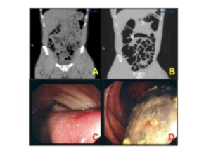
A 34 year old man was admitted to hospital with a history of progressively worsening abdominal pain associated with loss of weight. Clinical abdominal exam elicited left iliac fossa pain, but was otherwise normal. Blood tests were unremarkable. A CT KUB at presentation for suspected renal colic was performed and demonstrated a 5cm lesion in the descending colon involving the submucosa. A lumen filling lesion at the splenic flexure was confirmed at subsequent colonoscopy.
Laparoscopy was performed four months later and no masses associated with the colon were identified. An ‘on-table’ repeat colonoscopy identified no abnormalities other than an 8mm elevated area of mucosa in the distal transverse colon. A follow up CT virtual colonoscopy showed no evidence of a submucosal lesion. The patient currently remains asymptomatic.
A+B: side by side image of CT at admission and CTVC following on table colonoscopy
C+D: endoscopic images of stalk and of large necrotic lumen filling mass
Question:
What is the diagnosis and by what mechanism did the mass resolve?
Pomfret S1, Ahmed J1, Bhagat A2, Arbuckle J3, Landy J1.
1. Gastroenterology Department, West Hertfordshire Hospitals NHS Trust, U.K.
2. Department of Radiology, West Hertfordshire Hospitals NHS Trust, U.K.
3. Colorectal Surgery, West Hertfordshire Hospitals NHS Trust, U.K.
