In the May JNNP issue, Zhang and colleagues have published a study investigating the changes in structural and functional connectivity in patients with ALS.
ALS is one of the most complex neurodegenerative diseases, which affects the motor system. It is characterized by concomitant degeneration of the upper and lower motor neurons, producing progressive weakness and muscle atrophy. Even though it was initially conceived that just the motor cortex and anterior horn cells were affected, in recent years substantial evidence supports the compromise of extra-motor brain structures, such us the corpus callosum (CC).
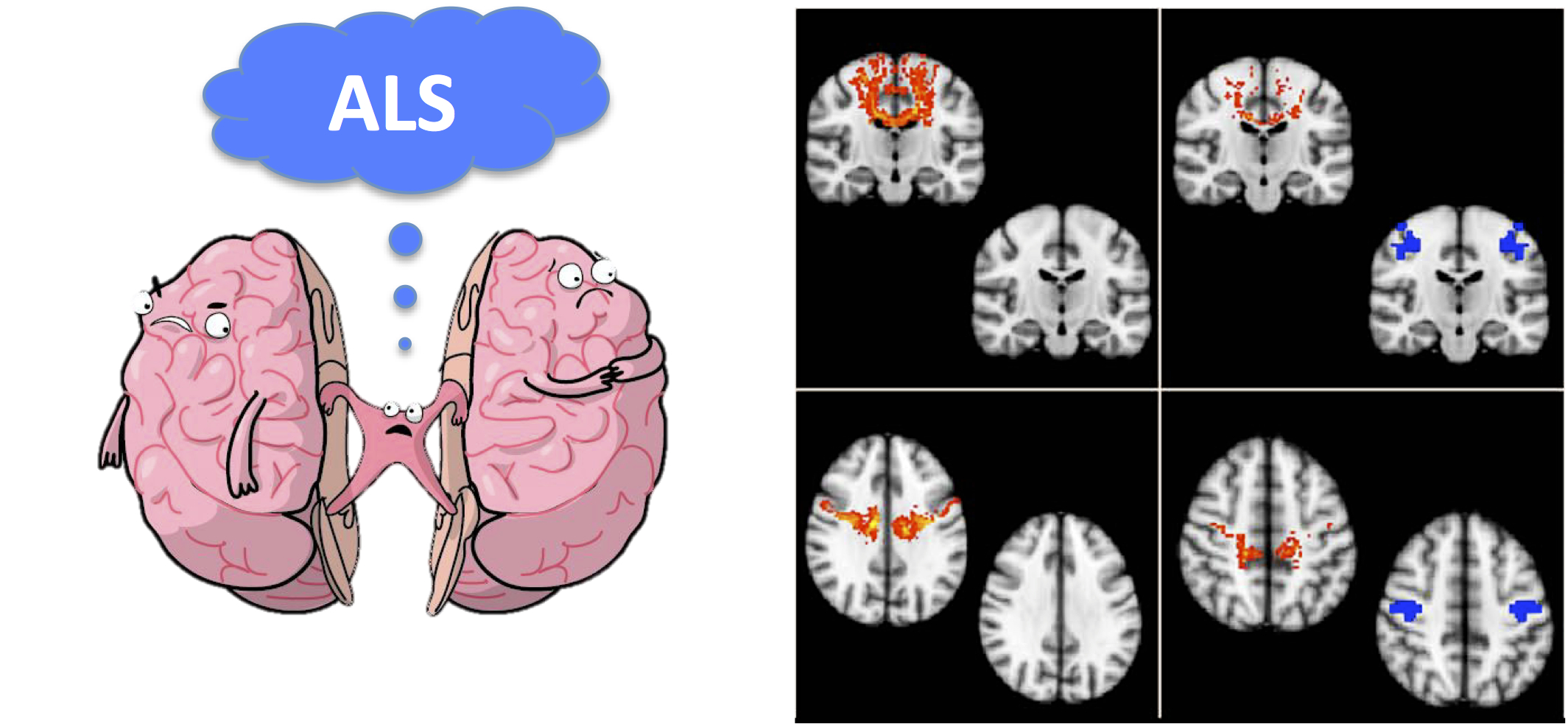
In this study, the authors have explored interhemispheric connectivity in 38 patients with ALS and 35 controls using diffusion tensor imaging (DTI) and resting state functional MRI (rfMRI). Indices of interhemispheric structural and functional neural connectivity were compared between groups. The rfMRI revealed a reduction in homotopic connectivity in ALS patients, specifically between the precentral and postcentral gyrus, the paracentral lobule, the superior temporal gyrus, the middle cingulate gyrus, the putamen and the superior parietal lobule, suggesting an extensive dysfunction in interhemispheric functional connectivity. In addition, DTI analysis in ALS patients showed a reduction in structural connectivity through the CC, specifically affecting subregions II, III and V. Finally, the combination of structural and functional data suggest that the central motor cortical interhemispheric fibres were the most affected in ALS.
This interesting article extends and confirms previous results that suggest dysfunction in interhemispheric communication, proposing a preferential involvement of the CC in ALS. Previous DTI studies in ALS have shown a reduction on fractional anisotropy in the CC extending to the primary motor cortex. On the other hand, electrophysiological studies have also supported the compromise of the CC. Specifically, transcranial magnetic stimulation (TMS) has provided evidence of functional impairment of the CC, showing a reduction in the interhemispheric inhibition. Clinically, a typical feature of ALS is focal clinical onset and regional spreading of neuronal degeneration. These clinical features indicate that neurodegeneration in ALS is an orderly propagating process, which seems to share the signature of a seeded self-propagation, such as prions. Interestingly, TDP-43, the major pathological protein in ALS, forms insoluble fibrillar aggregates in vitro and theoretically can act as seeds to trigger the aggregation of native proteins. This cumulative evidence allows us to question the contribution of the CC in spreading of disease and its potential use as a therapeutic target in the future.
Read more at http://jnnp.bmj.com/content/88/5/369.1