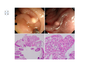
An 82-year-old woman with a history of eosinophilic granulomatosis with polyangiitis treated with 4 mg/day prednisolone underwent a colonoscopy because of a positive fecal occult blood test. This revealed a 2-mm slightly elevated yellowish lesion in the transverse colon (figure 1). Narrow-band imaging showed intact pits of the colonic mucosa (figure 2). Physical examination was unremarkable. Laboratory test results, including triglyceride and cholesterol levels, were also unremarkable. A biopsy was taken (figure 3, 4).
Whats the diagnosis?
KentaHamada1,2, Yasushi Yamasaki1, Jun-ichi Kubota2, Hiroyuki Okada1
1Department of Gastroenterology and Hepatology, Okayama University Graduate School of Medicine, Dentistry and Pharmaceutical Sciences, Okayama, Japan
2Department of Internal Medicine, Tajiri Hospital, Mimasaka, Japan
