Professor El-Omar has chosen Dr Mario Dinis-Ribeiro, CIDES/CINTESIS, Faculty of Medicine, University of Porto, Porto, Portugal, and Dr Cesare Hassan, Digestive Endoscopy Unit, Nuovo Regina Margherita Hospital, Rome, Italy, to do the next #GUTBlog.
The #GUTBlog focusses on the latest paper “Standalone performance of artificial intelligence for upper GI neoplasia: a meta-analysis” which was published in paper copy in GUT in August 2021. Dr Dinis-Ribeiro and Dr Hassan are the senior authors on this paper.
Dr Mario Dinis-Ribeiro and Dr Cesare Hassan write:
“One out of 4 European citizens died of cancer in 2016, ranking second in the causes of death in Europe. The EU Mission for Cancer was established and aims at, in 2030, 3 millions of lives being saved, live longer and better.
Digestive cancers represent a significant proportion of these with a particular feature that is the chance for early diagnosis namely by non-invasive approaches (e.g. FIT for colorectal cancer) or digestive endoscopy. However, for colorectal cancer, most countries have or are planning to have established population-based screening programmes with clear long-term benefits, upper GI neoplasms (e.g. oesophageal squamous and adenocarcinoma and gastric adenocarcinoma) lack evidence to suggest similar approaches.
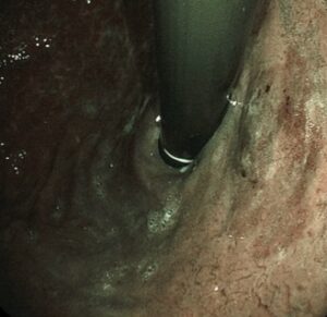
Therefore, early diagnosis during an upper gastrointestinal endoscopy with a case-by-case approach represents a unique chance to achieve the goal of minimal invasive approach, improving survival and quality of life. However, missing rates are not negligible due to the subtle appearance, heterogeneous pattern across different types of upper-GI cancer (squamous versus Barrett-related versus gastric cancer), and lack of proper training at least for Western endoscopists.
When performing an Upper GI (UGI) endoscopy in daily practice, we believe we are offering a unique gold standard for cancer diagnosis, but almost paradoxically the chance of missing neoplasia, especially in an early stage, is far from being zero! When considering a 10-20% miss rate, the loss in terms of life-years due to false negatives at upper-GI endoscopy is substantial, especially when considering the steep worsening of prognosis with cancer upstaging.
Artificial intelligence (AI) based on object-recognition is already impacting our daily working lives and AI already deeply penetrated in the field of colonoscopy. On the other hand, incorporation of AI for upper-GI endoscopy appears to be more problematic due to the development of disease-related software, preventing a synoptic view of AI development. That is, we were missing “the” summary defined as the current status of what has been produced by our endoscopic researchers in the field of AI and upper GI endoscopy and how can determine is current role and impact.
In our manuscript, we provided a synoptic estimate of the accuracy of all the AI that were developed for detection of upper-GI neoplasia, including squamous cell, Barrett-related neoplasia, and early gastric cancer. Such estimates were derived by standalone performance studies where the AI software was validated against images of upper-GI neoplasia detected by expert endoscopists in an artificial setting. Overall, we showed an impressive 90% sensitivity of AI for neoplasia detection with an overall area-under-the curve of 0.95. In addition, we showed no difference in AI performance across the three main types of upper-GI neoplasia, showing a categorical difference with the human experiences that is heavily biased by the type of neoplasia included.
These data strongly support the simultaneous implementation of a global AI system for all the possible neoplastic lesions detectable by AI for its clinical relevance. This will minimize the miss rate of upper-GI neoplasia detection, irrespectively of the patient or endoscopist profile. When looking at the correspondence between standalone performance and clinical outcome of AI in colonoscopy, there is no reason why such good data should not prompt better detection rates at clinical level. Whether this involves the creation of only one global software or the grouping of different software in the same machine may be considered as a technical rather than clinical barrier.
What should be addressed in the next future is the impact of AI on the clinical outcome of upper-GI neoplasia. This depends on several factors, such as the interaction between human endoscopist and machine in vivo. Will the endoscopist accept as true positive all the neoplastic cases detected by AI? Will the AI-induced false positives trigger useless resection or generate anxiety? Secondly, will Western endoscopists learn the proper characterization phase for these lesions, such as the intrapapillary capillary loops classification for squamous cell neoplasia or the PREDICT for Barrett-elated neoplasia?
In other words, our synoptic summary prompts a quick implementation of AI in upper-GI endoscopy but even a more immediate analysis of its clinical benefits and possible drawbacks.”
Dr Mario Dinis-Ribeiro and Dr Cesare Hassan