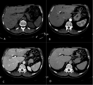
Figure 1
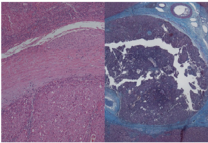
Figure 2
A 62 year old male was undergoing antiviral therapy for HCV cirrhosis. He was asymptomatic with normal liver function tests and a normal alpha fetoprotein level. Routine liver ultrasound suggested a new portal vein thrombosis. CT imaging (figure1) and subsequent pathology specimen (figure 2) demonstrate a unique lesion. What’s in the portal vein?
Submitted by
J.Doherty, C.Braniff, S.Oon, J. O’ Neill, P A. Mc Cormick.
Liver transplant Unit, St Vincent’s University Hospital, Dublin.
(Visited 636 times, 1 visits today)

